Role:
Illustration
Tools:
Procreate, Illustrator, InDesign
Date:
Mar 2024
Publication:
The University of Tennessee College of Medicine Chattanooga's Department of Orthopaedic Surgery performed a novel repair technique using knotless, all-suture anchor technology intended for higher-grade, operative AC joint injuries in “high-risk” patients, i.e., those returning to a collision sport such as football, rugby, hockey, or wrestling. In addition, this technique could be employed in patients at risk for delayed or nonhealing, such as those with diabetes or who are smokers, those at risk of noncompliance, and revision cases. The all-suture anchor, knotless “suture staple” technique can be implemented easily to provide backup fixation of the AC joint directly as an augmentation to CC reconstruction, preferably arthroscopic-assisted reduction, and fixation with a cortical button and, when indicated, concomitant allograft reconstruction.
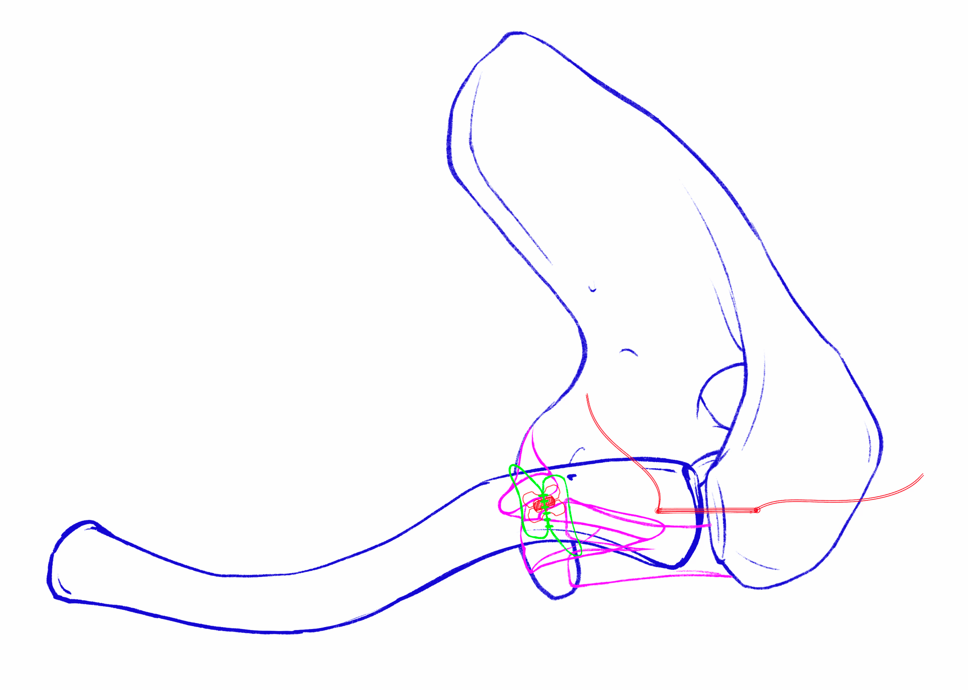
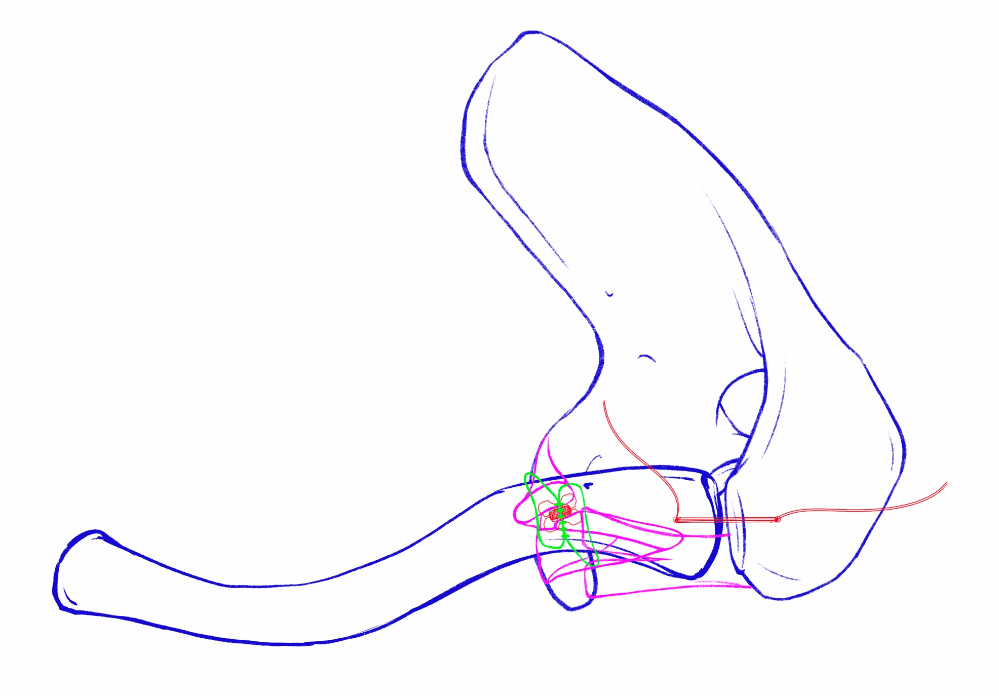
Since visualization of this area is difficult even with surgical video and cadaver demonstration, I was asked to prepare illustrations for publication of their findings.

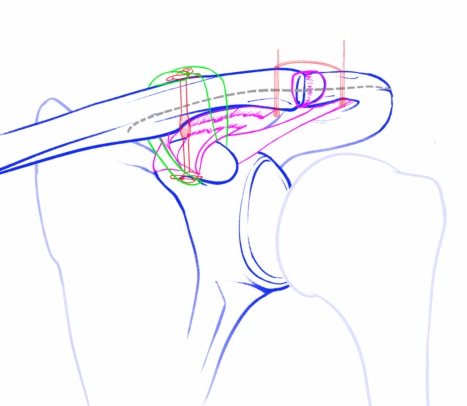
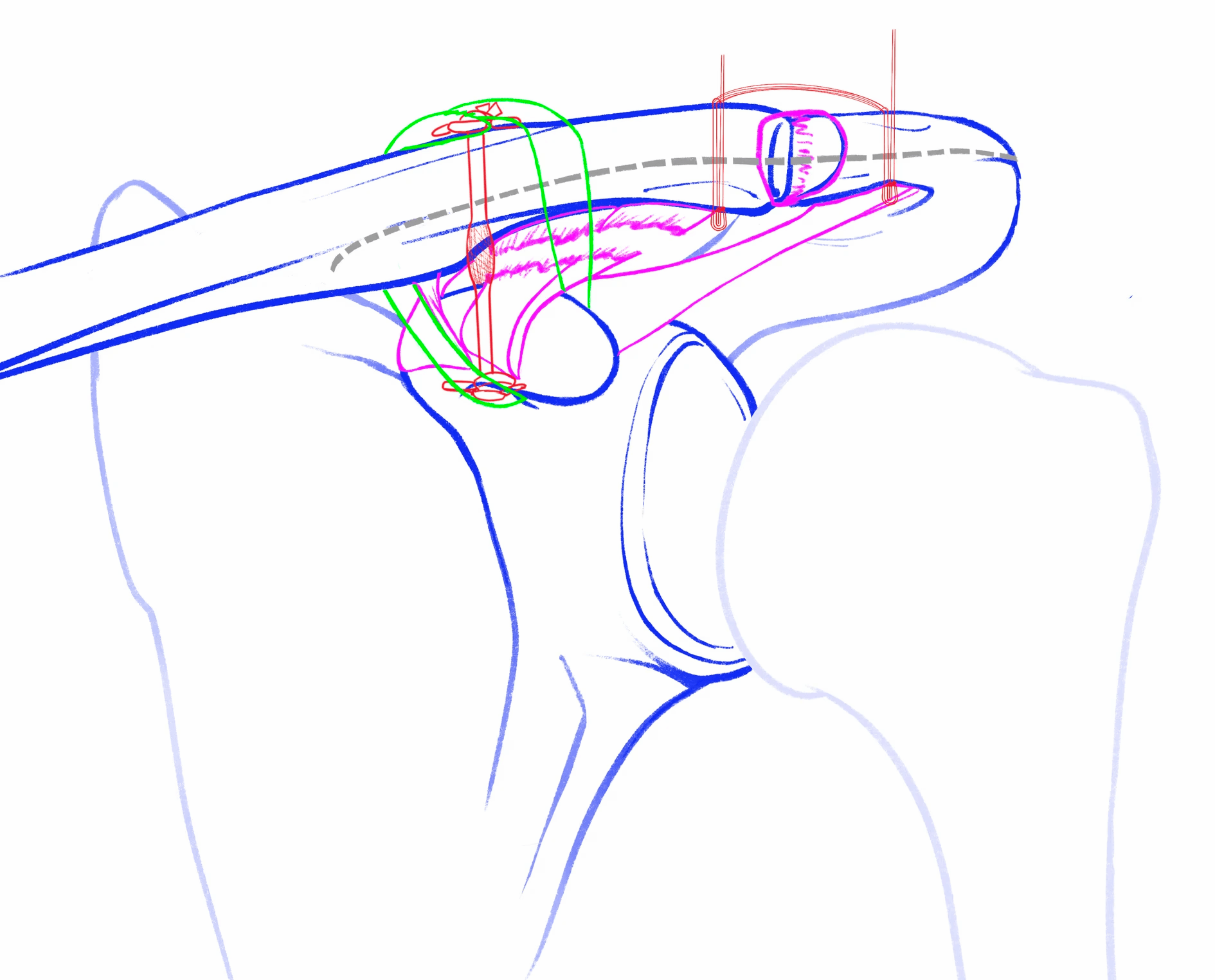
Fig 5. (1) Illustrated anterior view of the final construct showing the complete coracoclavicular fixation with Dog Bone suspensory device and looped gracilis allograft with anterior and posterior limbs sutured together. (2) Illustrated superior view of the construct again demonstrating the completed coracoclavicular fixation with Dog Bone and gracilis allograft as well as the suture staple across the AC joint before cutting of the excess suture limbs.
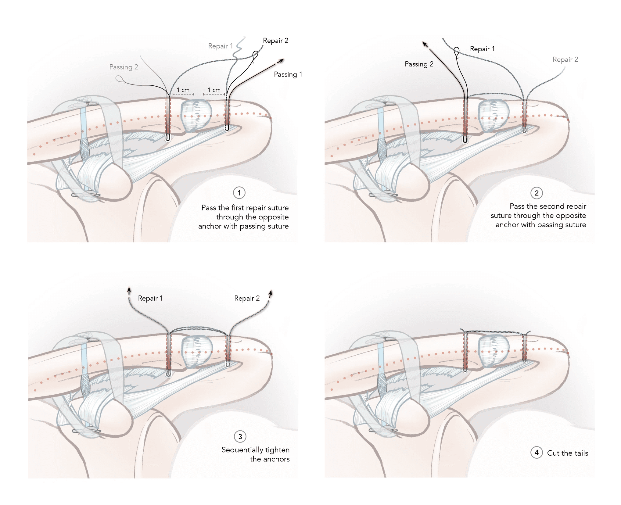
Fig 2. (1) Two 1.8-mm knotless, all-suture anchors are placed approximately 1 cm from the acromioclavicular joint, with care taken not to penetrate the joint or anterior/posterior cortex when drilling. After anchor placement, proceed with suture passing by shuttling the repair stitch through the opposite anchor’s passing suture. (2) Repeat suture passage for the opposite anchor. Passing can be completed beginning with either acromial or the clavicular anchor. (3) Using gentle, sequential pulls on the tails, secure the construct until satisfactory tension. (4) Cut the remaining tails once final tension has been achieved to complete the knotless construct.
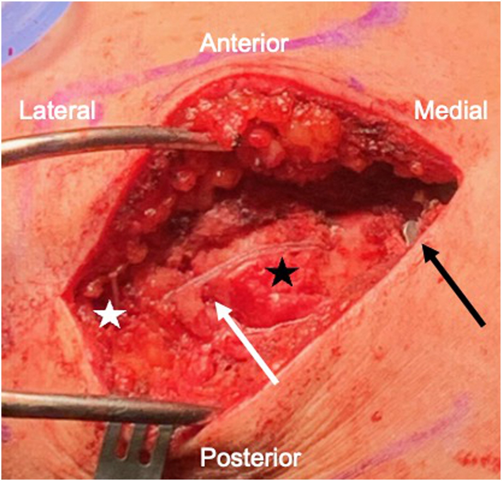
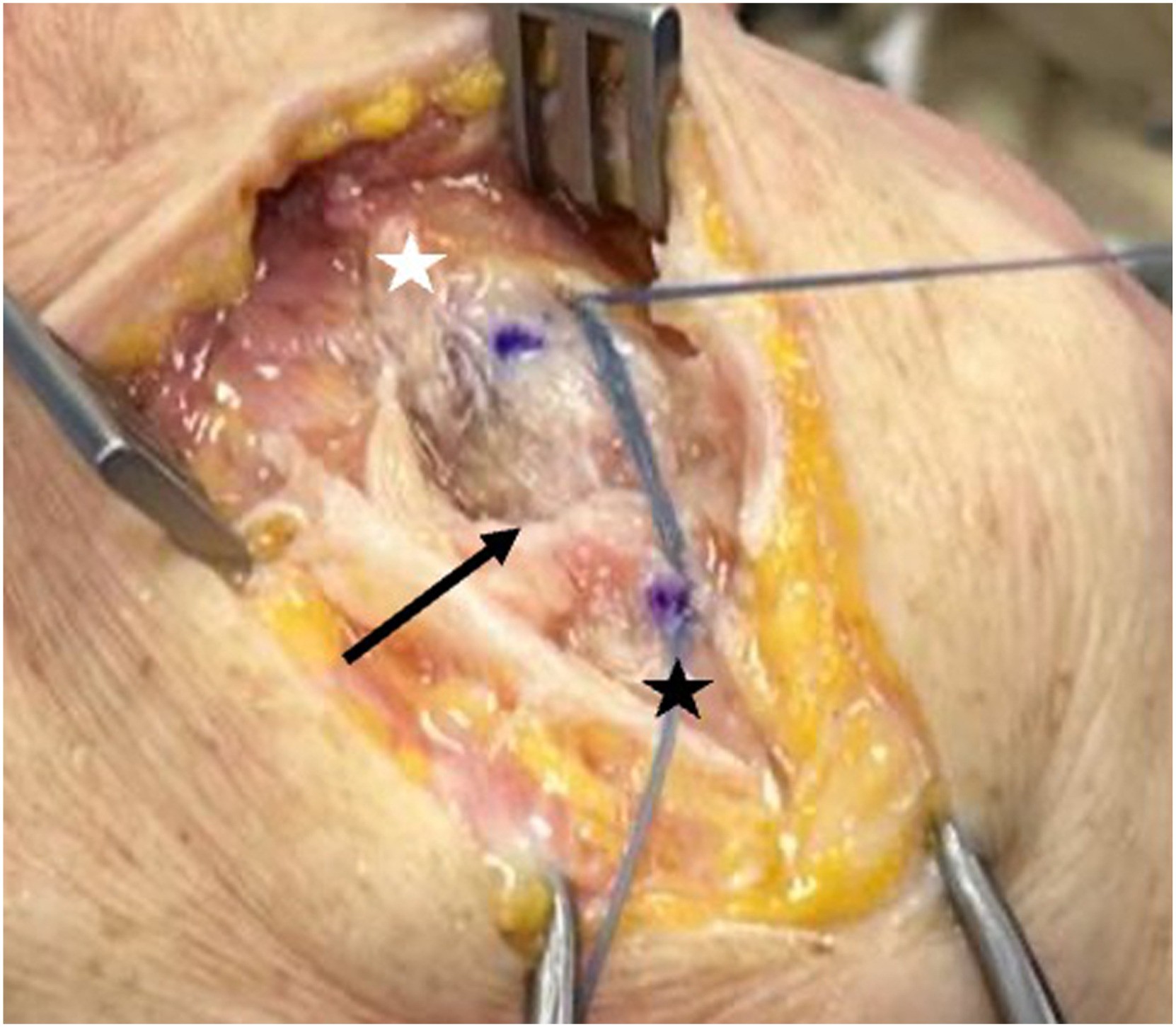
Fig 4. Open, superior view of the acromioclavicular joint (black arrow) demonstrating the suture staple configuration linking the distal clavicle (white star) and acromion (black star), after repair suture shuttling through the adjacent anchor and preliminary tightening.

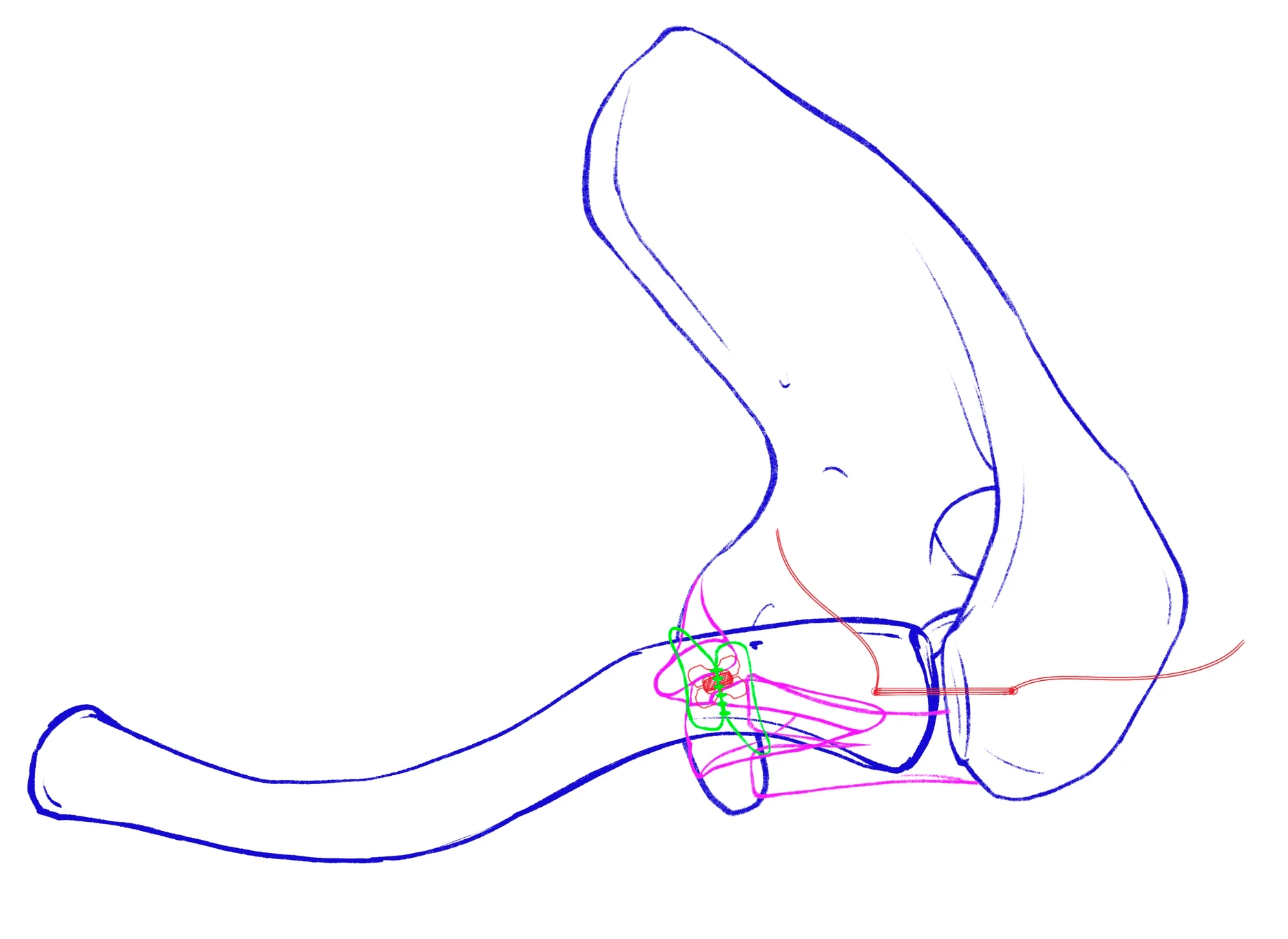
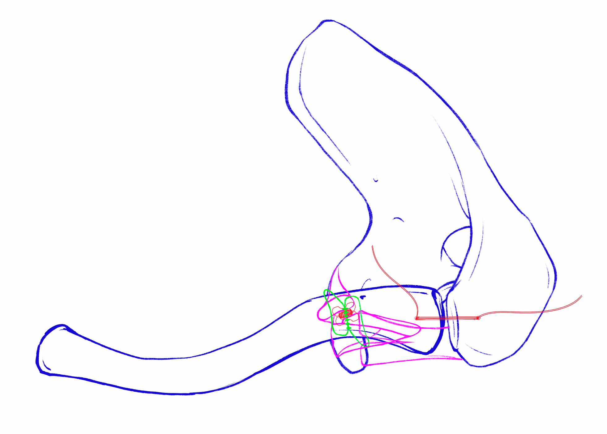
Consultation sketch by Dr. White


R E F E R E N C E S